

Practice Perfect 830
Making Practice Perfect Decisions
Part 7 - Know Where You Are
Making Practice Perfect Decisions
Part 7 - Know Where You Are
In today’s issue of our continuing series Making Practice Perfect Decisions, we are going to get a little granular and detail oriented. The principle up for discussion today is to:

This principle is really about optimizing the factors to make a surgical procedure successful. Anyone trained in surgery has had moments where they’ve had to fight with a poorly placed incision or a patient that is positioned in such a way as to make the procedure more challenging.
Today we’re going to discuss the following:
- 3D visuospatial knowledge
- Patient positioning
- Incisional approach
- At risk structures
- Open it so you can close it
- Tissue preservation
3D Visuospatial Knowledge
It may seem obvious that a primary requirement of doing any surgery successfully is having a solid understanding of the three-dimensional nature of the lower extremity. However, in my experience working with trainees, they oftentimes lose their sense of location. For example, placing a guide pin for an Austin bunionectomy in the proper direction requires an understanding of the angle of the pin in reference to the metatarsal but also appreciating where the foot is sitting in space. If the foot is externally rotated on the surgical table, and the trainee does not appreciate its position, that guide pin may not be placed in the proper trajectory. Similarly, when performing dissections for soft tissue masses, it is easy to become lost when starting to remove the mass itself. Maintaining an understanding of three-dimensional location is imperative.
Patient Positioning
This is an often underappreciated yet highly important aspect of preparing the patient for surgery. As a simple example, it is rare to repair a calcaneal fracture with the patient supine and the leg externally rotated. This would make it very difficult to access the lateral side of the heel. Additionally, when using intraoperative fluoroscopy, the other foot may get in the way and show up on the image if the foot is not properly positioned. My personal preference is to place the foot at 90 degrees to the surgical table in almost all supine cases and, when using a fluoroscope, placing blankets under the surgical limb to elevate it so that lateral views do not involve the other foot. Similarly, I like to have the foot right at the edge of the bed and occasionally with the heel hanging off depending on the procedure. In other cases, some combination of these positions may be appropriate with some extra attention to detail. For example, when performing a posterolateral approach to an ankle fracture, having the patient in a more lateral position but placing the bean bag in such a way as to allow the leg to externally rotate makes it possible to repair a medial malleolar fracture at the same time without having to reposition the patient. Every little detail counts.
Incisional Approach
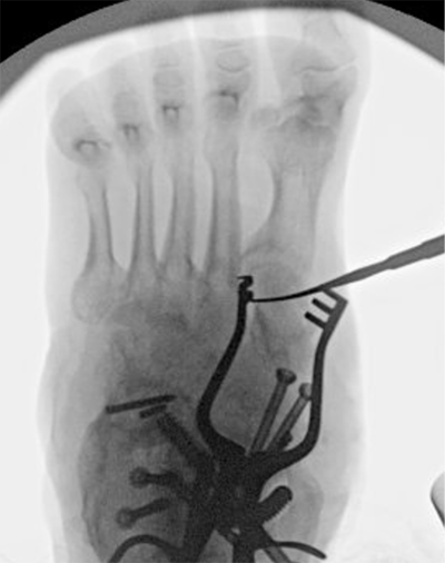
This is obviously a very important concept. Placing the incision in the wrong location can be detrimental to performing a procedure efficiently. It is worth taking a few extra minutes to make sure that an incision is placed properly. For example, Figure 1 shows the use of fluoroscopy and a freer elevator to find the location of the medial cuneiform for a cotton osteotomy. Now, it is possible to create this incision by palpating the anatomy, but an extra moment to double check the location can allow one to create a smaller incision.
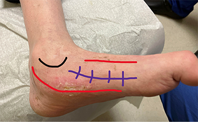
It is also important to remain cognizant of the surface anatomy to help one create proper incisions. As an example, as shown in Figure 2, palpating the dorsal and plantar aspect of the medial column allows one to make a central incision for surgical access to this area. There is often a tendency to accidentally make this incision too far plantar during medial arch procedures. Again, carefully palpating the area and obtaining intra operative fluoroscopy views can help create optimal access to the surgical area and an easier procedure.
At-Risk Structures
For experienced foot and ankle surgeons, this is deeply ingrained in our DNA. We are constantly on the lookout for important anatomy that is easy to damage. For newer surgeons, it is helpful to quickly run through in your mind the important anatomy and carefully protect those structures during the procedure. Consideration of these at-risk structures is also imperative when determining incision location.
Open It So You Can Close It
This phrase was introduced to me by one of my residency partners, an excellent surgeon. He likes to say that it is easy to open the foot, but, without proper dissection, it is often difficult to close layers. While making an incision and beginning the dissection, consider how you will close that area once the procedure is over. For example, when performing a Lapidus bunionectomy or a metatarsal head osteotomy, I typically incise approximately five millimeters medial to the extensor digitorum longus tendon to leave a cuff of tissue attached to the extensor tendon. This will allow closure of that layer with ease.


Similarly, consider that most dissection of the foot and ankle has a consistent pattern where the surgeon incises the skin, spreads the subcutaneous layer atraumatically with a scissor - simultaneously obtaining hemostasis - then returns to the use of a scalpel for the facial layers, and may use a variety of elevators to tease off the periosteum. This careful dissection permits an easy layered closure. Keeping this concept in mind will allow one to maintain an organized approach to dissection, no matter what part of the foot or ankle you are working.
Tissue Preservation
Everything we have discussed really boils down to preserving the patient’s tissue without causing excessive damage whenever possible. Obviously, sometimes it is necessary to create damage to help the patient but maintaining as much normal anatomy as possible is pivotal to obtaining the best result possible. As an example, I am usually not trying to make the tiniest incision for an Austin bunionectomy because fighting with a very small incision may create damage to the skin. The classic statement incisions heal side to side and not end to end is very true. Now, this does not eliminate the possibility of performing minimal incision surgery in the proper patient and procedure, but patient well-being should never be compromised in order to make a smaller incision.


Knowing where you are, then, contains many important principles that will help make for successful surgery and happy patients.
Best wishes.

Jarrod Shapiro, DPM
PRESENT Practice Perfect Editor
[email protected]

Major Sponsor


Comments
There are 0 comments for this article