
Practice Perfect 979
Dermoscopy: A New Instrument for Podiatrists?
Part 1: What Is It?
Dermoscopy: A New Instrument for Podiatrists?
Part 1: What Is It?
Recently, I was turned on to a technology that I had never heard of for most of my practice life: dermoscopy. This is a very common instrument in dermatology circles, but I’d never seen it used in podiatry. After investigating it further, I’ve found it to be a promising technology for podiatrists that I’d like to share today. Let’s review this tech and provide a few examples where it can be used in podiatric medicine. Before we get started, I must claim that I am a novice with dermoscopy, and for those interested, I strongly recommend doing your own study.
What is Dermoscopy?
This technology uses a device called a dermatoscope to visualize lesions of the skin. In dermatology, this is primarily used to examine pigmented lesions of the skin, but there are several uses and expanding indications. For example, this may be used to examine the nail unit (see below), which is called onychoscopy. Similarly, one can look for carcinomas, melanoma, and other disorders, to differentiate them from benign skin conditions.

Dermatoscopes use magnification and polarized and nonpolarized light to both better view skin lesions and to examine specific characteristics of those lesions. This is not a glorified magnifying glass. Because of the way light reflects off of skin, we are not normally able to see deeper structures. The two different types of light from the device allow additional features to be seen that are not normally easily visible to the human eye. The polarized setting allows indirect contact (the device does not touch the patient), while using the nonpolarized light setting requires a contact medium such as ultrasound gel.
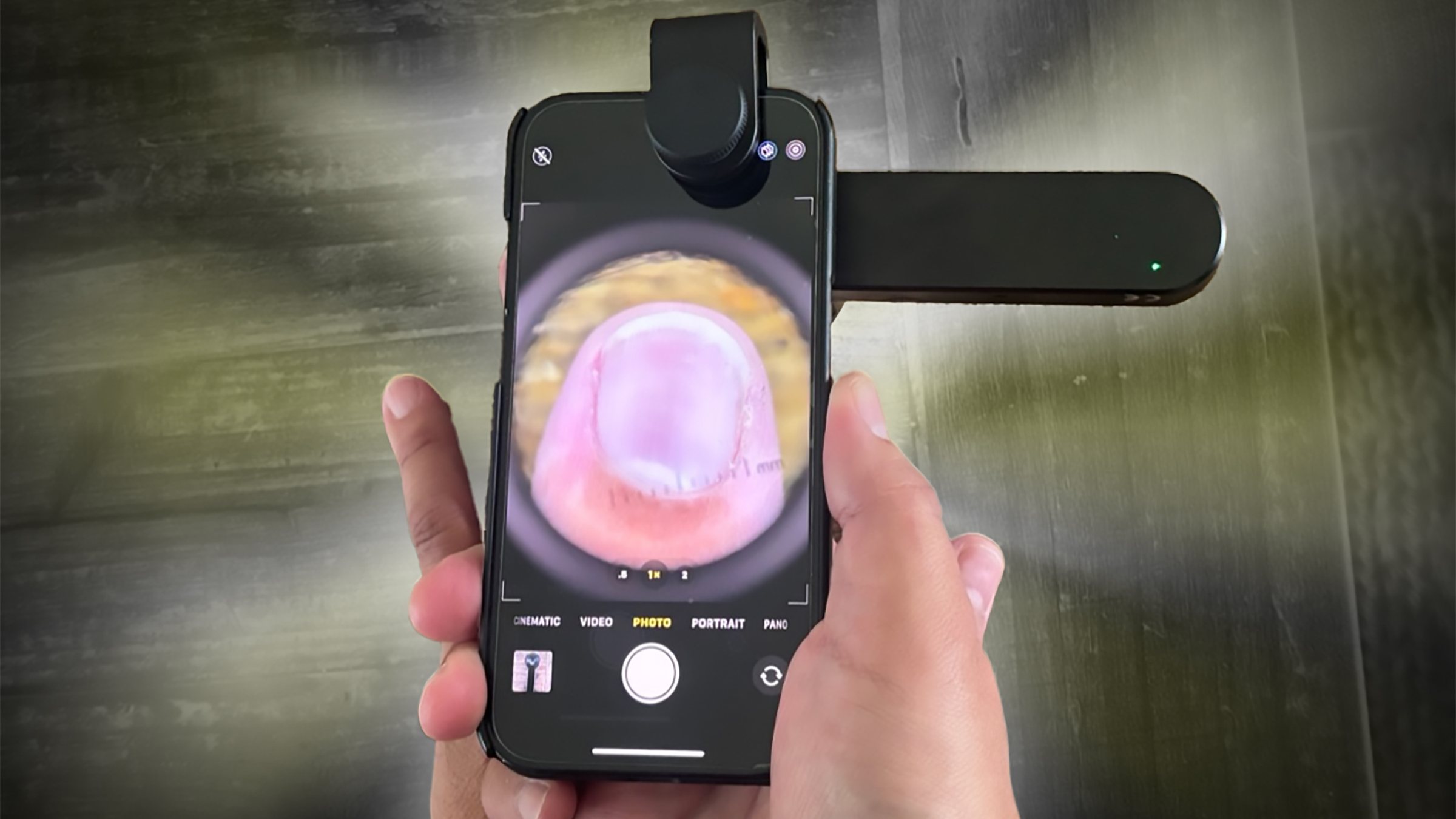
Figure 1 shows an example of a dermatoscope. In the past, the physician would use the dermatoscope by itself, looking straight into the skin. Today, newer devices allow attachment to a smart phone, and simply using the camera app allows one to easily see the image and keep those images for documentation purposes. The dermatoscope itself attaches via a magnet to the phone, as shown in Figure 1. There are a few different devices that may be purchased, but the one I chose was an Iboolo DE-3100 hybrid dermatoscope (it has both polarized and nonpolarized options), and it cost $490. It takes about 30 seconds to set up and is easily used in the podiatric clinic.
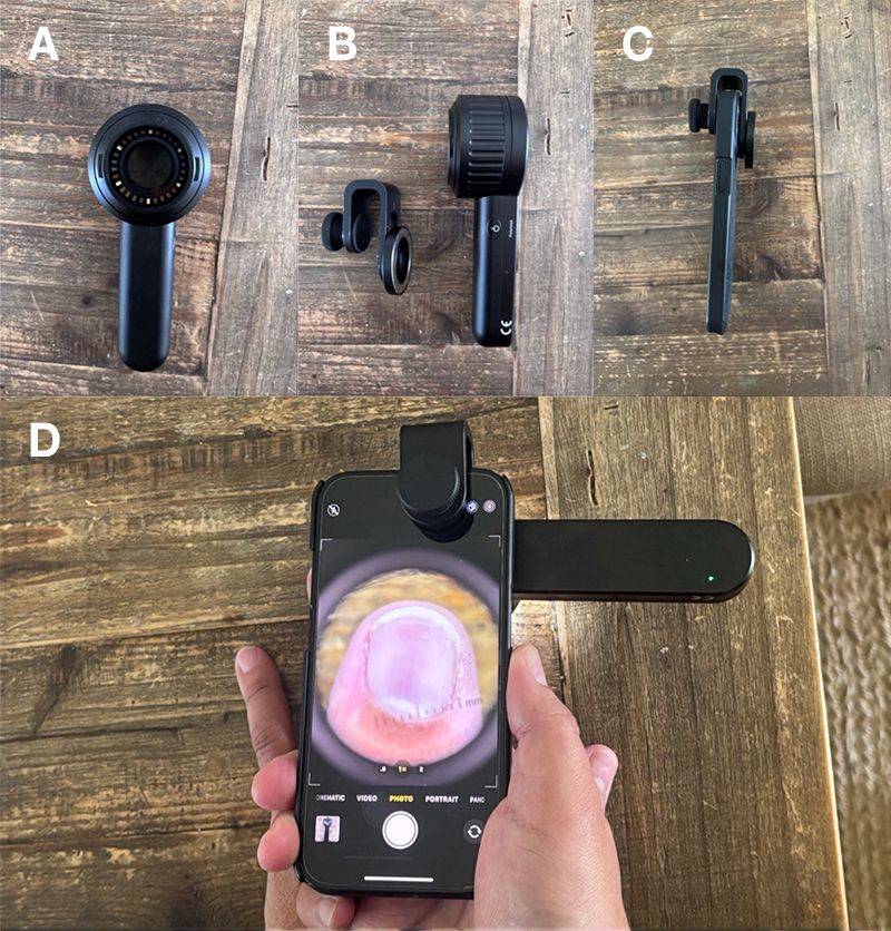
Figure 1. Example of dermatoscope. A = en face view of the device.
B = Dermatoscope + smart phone mounting device. Polarized light/power button shown. Nonpolarized button on opposite side. C = Smart phone with mounting device attached. D = Entire unit powered on showing the author’s index fingernail at 10X magnification using polarized light.


Is This a “Plug and Play” Technology?
Yes and no. The devices themselves are very easy to use, rapid, and intuitive. Using it is the easy part. The more complicated part is the image interpretation in an expert manner. Dermoscopy utilizes pattern recognition and a strong understanding of the anatomy and physiology of the skin to truly understand what one is seeing in the image. For example, pigmented lesions that are in the dermoepidermal junction of the skin tend to appear slightly darker brown, while melanin in the stratum corneum of the epidermis will appear much darker, possibly black. Similarly, onychomycosis is diagnosed via a pattern of destruction of the nail by seeing longitudinal white striae, a jagged proximal edge, and subungual hyperkeratosis in a “ruin” pattern. By successfully combining knowledge of skin structure anatomy, pattern recognition, and the regular history and physical examination, one may build a comprehensively obtained diagnosis.
Does This Replace Biopsy?
Absolutely not. Biopsy is still the gold standard for pathologic diagnosis of skin structure lesions. Think of dermoscopy in the same way you might regard advanced imaging. The physician sees a patient, does a history and physical, obtains radiographs, and sometimes uses MRI for more anatomical information. By combining all this information, one may arrive at a better diagnosis. The same is true for dermoscopy.
Can I Bill for Dermoscopy?
There are a few CPT codes that dermatologists use, but my own investigation of these codes does not show they translate into regular podiatric use. For those interested, I suggest referring this question to coding experts.
How Can I Learn More?
For anyone learning to use this technology, here are some suggestions. Amazon has a number of textbooks ranging in cost from about $30 to about $300. I can’t believe I’m writing this, but there are actually a few very good resources on YouTube. Check out Primary Care Dermoscopy Academy for an excellent education set up almost like a med school course (but more engaging). There are also several medical research studies on the use of dermoscopy as well covering various important clinical topics.

To wrap up this introduction to dermoscopy, this technology has the potential to help us make stronger dermatological inferences for better decisions. In next week’s Part 2 we’ll discuss a few of my initial cases to get a feel for where this can be used in the lower extremity.
Until then, best wishes.

Jarrod Shapiro, DPM
PRESENT Practice Perfect Editor
[email protected]

This blog and the overall sponsorship of PRESENT Podiatry was made possible through the support of our sponsors:


Comments
There are 0 comments for this article