

Xerosis Cutis

Expert Opinion by
Beau DiCicco, MD, FAAD
Introduction: Xerosis cutis is the diagnostic term for dry skin. Dry skin can be caused by either exogenous factors or endogenous factors.1
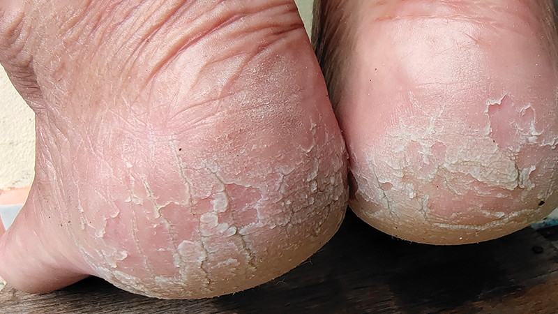
Patient Impact: Xerosis cutis can impact patients both physically and psychologically. Patients often experience painful symptoms like dryness, itchiness, and skin cracking, all of which can lead to psychological discomfort due to the unsightly appearance of the condition.1,2
Clinical Outline:
- Epidemiology
- Xerosis cutis is a common skin disorder, yet most patients tend to self-treat this condition at home, leading to an underreported prevalence of the condition.
- Etiology
- The lower level of the stratum corneum is composed of corneocytes and filaggrin, which maintain hydration.
- The upper level of the stratum corneum is composed of lipid agents that protect the skin from harmful agents and bacteria.
- Diagnostics
- Diagnosis of xerosis cutis is clinically based.
- There are other methods available to the physician to measure the roughness, sebum production, and amount of water loss.
- Treatment
- Based on the presentation of symptoms, the treatment formulation should include a combination of moisturizers, lipid replenishing agents, and skin soothing agents.
Expert Opinion with Beau DiCicco, MD, FAAD:
On the discussion with patients regarding xerosis cutis…
I inform my patients that this is a common condition, especially during the winter months when heating systems dry the air. Additionally, I set expectations regarding treatment. Xerosis cutis is a chronic condition that fluctuates based on several factors including the weather, stress, and comorbidities. Patients need to understand that there is no single treatment to “cure” this dermatologic condition. It requires long term treatment.
On the treatment of xerosis cutis…
My initial treatment involves either 40% urea cream or ammonium lactate. I sometimes add lipid replenishing agents, such as ceramides, and skin soothing agents to the prescribed treatment, though these are not always necessary. I advise patients to apply the cream to the dry areas of their feet and immediately cover them with socks. While I would prefer my patients to do this throughout the day, many will find it uncomfortable, so I recommend applying it at least every night.


Epidemiology: There is no gender predilection for xerosis cutis.3 However, prevalence does increase with age. Xerosis cutis as a distinct diagnosis is underreported compared to its actual prevalence, as this condition is often self-treated.1,4
Additionally, xerosis cutis is a common secondary condition from autonomic neuropathy. In patients with autonomic neuropathy the nerves that control sweating is damaged.
Etiology: Keratinocytes are the predominant cells of the epidermis, originating from the basal layer. These cells differentiate into corneocytes, which migrate from the basal membrane to the skin surface over a period of approximately 4 weeks. During this process, corneocytes differentiate into enucleated, organelle-free cells surrounded by a rigid envelope that eventually sheds off. Corneocytes enhance the mechanical strength of the epidermis, enabling this skin layer to act as a physical barrier. This barrier prevents the entry of harmful agents and helps the body retain moisture. Additionally, profilaggrin, a protein in the epidermis, converts into filaggrin, which facilitates the formation of disulfide bridges between keratin filaments in the stratum corneum. These bridges provide an additional protective layer, similar in function to the corneocytes, further reinforcing the skin’s defensive capabilities.1
Above the corneal layer is the intercellular lipid bilayer, composed of keratinosomes (Odland bodies), which are mixtures of ceramides, sterols, and other free fatty acids. Additionally, when filaggrin is degraded into pyrrolidine carboxylic acid, urocanic acid, and free amino acids, these components are converted into natural moisturizing factors (NMF). NMFs are essential humectants that aid in water retention. Furthermore, there is a thin hydrolipid layer consisting of lipids produced by sebaceous glands, sweat, and remnants of keratinocytes that are sloughed off. This layer provides added protection against harmful external agents. 1
Although this model of the skin is simplified, it provides valuable information about the structures responsible for the development of xerosis cutis. Xerosis is caused by damage to the superficial lipid bilayer, altered keratinocyte differentiation, or a decreased presence of NMFs. This etiology can be attributed to either exogenous or endogenous factors.

Xerosis can be caused by exogenous factors such as environmental conditions (cold, low humidity), occupation (employment in fields that exposes patients to irritants), or cleansing patterns (frequent long hot showers, use of soapy irritants). Endogenous factors that result in xerosis cutis include inflammatory skin disorders (atopic dermatitis, psoriasis), endocrine disorders (diabetes, hyperparathyroidism), or prescribed medication (retinoids, topical corticosteroids).
Diagnostics: The diagnosis of xerosis cutis is based on physical exam findings. Several grading scales are available for xerosis cutis, including the Overall Dry Skin Score (ODS), that assesses the presence of scaling, roughness, redness, and cracks (SRCC), and the diagnostic xerosimeter.1,5 To further detail the pathologic skin, certain diagnostic tools are available. The amount of transepidermal water loss (TEWL) can be measured using tewametry.6 Skin hydration can be measured through cornometry. 1
Treatment: The goal of skin care of xerosis cutis is to improve skin hydration and compensate for the lack of the lipid barrier. Before prescribing medication to treat xerosis cutis, it is important to discuss preventative measures. These measures include the use of gentle cleansing agents, changing clothing to cotton, and dietary adjustments such as avoiding citrus and spicy foods. Additionally, patients should avoid hot, dry environments and excess sun exposure.1,7,8 Once preventative measures have been exhausted, prescribed skin care agents should include a mixture of moisturizers (eg, urea, lactic acid derivatives, and glycerol), lipid replenishing agents (ceramides, natural oils), and skin soothing agents (vitamins).
Moisturizers: Of all moisturizers, urea is considered the gold standard in the treatment of xerosis cutis. Urea, an NMF, hydrates the skin and improves corneal barrier strength. It acts as a humectant, which increases hydrogen bond formation with non-polar aromatic amino acids, which increases the water binding capacity of the corneal layer. Additionally, urea can modulate the expression of genes responsible for lipid synthesis and keratinocyte differentiation.9 Specifically, urea concentrations between 10-25% are associated with effective treatment of xerosis cutis of the feet.
Ammonium lactate, which contains lactic acid, is another moisturizer that includes NMF. Although the function of ammonium lactate when treating xerosis cutis is similar to that of urea, urea 40% is superior in efficacy compared to 12% ammonium lactate.10
Glycerol, another moisturizing agent, increases the number of aquaporin-3 channels in the skin, thereby increasing the ability of the stratum corneum to retain water. Glycerol concentration in prescribed lotions can be up to 20%, but are typically found in concentrations between 5-10%.1,11

Lipid Replenishing Agents: Ceramides, which are sphingolipids, are lipophilic agents essential to the lipid barrier. Ceramides make up approximately 40% of the extracellular lipid matrix in the stratum corneum which protects the skin from harmful invading agents. It is essential for products containing ceramides to be in a physiological lipid composition that includes fatty acids and sterols.1,11
Natural oils are various substances that differ in their content of triglycerides, free fatty acids (omega-3, omega-6, and saturated fatty acids), and mono saturated oleic acids. These oils act as a hydrophobic protective layer on stratum corneum, thereby providing a protective barrier against invading agents.1,16
Skin Soothing Agents: A product containing skin-soothing agents, such as Licochalcone A, oat extract, and dexpanthenol, is used in severe cases of xerosis cutis. Licochalcone A, a derivative of the licorice plant, and oat extract inhibit the section of pro-inflammatory factors, which relieve patient discomfort.13,14
Dexpanthenol, a precursor of vitamin B5, reduces oxidative damage by stimulating the production of antioxidant, providing further relief to patient discomfort.15
Conclusion: Xerosis cutis is caused by either exogenous or endogenous factors. Exogenous factors include environmental influences, occupational exposure, and patient cleansing habits. Endogenous factors encompass inflammatory skin disorders and prescribed medications. Xerosis cutis results from damage to the intercellular lipid bilayer, altered keratinocyte differentiation, and decreased moisturizing factors on the skin. The diagnosis of xerosis cutis is based on clinical evaluation. Treatment of xerosis cutis involves a combination of moisturizers, lipid replenishing agents, and skin soothing agents.

- Augustin M, Wilsmann-Theis D, Körber A, et al. Diagnosis and treatment of xerosis cutis - a position paper. J Dtsch Dermatol Ges. 2019 Nov;17 Suppl 7:3-33. doi:10.1111/ddg.13906
Follow this link
- Tuckman A. The Potential Psychological Impact of Skin Conditions. Dermatol Ther (Heidelb). 2017 Jan;7(Suppl 1):53-57. doi:10.1007/s13555-016-0169-7
Follow this link
- Augustin M, Kirsten N, Körber A, et al. Prevalence, predictors and comorbidity of dry skin in the general population. J Eur Acad Dermatol Venereol. 2019;33(1):147-150. doi:10.1111/jdv.15157
Follow this link
- Thyssen JP, Johansen JD, Zachariae C, Menné T, Linneberg A. Xerosis is associated with atopic dermatitis, hand eczema and contact sensitization independent of filaggrin gene mutations. Acta Derm Venereol. 2013 Jul 6;93(4):406-410. doi:10.2340/00015555-1539
Follow this link
- Serup J. EEMCO guidance for the assessment of dry skin (xerosis) and ichthyosis: clinical scoring systems. Skin Res Technol. 1995 Aug;1(3):109-114. doi:10.1111/j.1600-0846.1995.tb00029.
Follow this link
- Ali SM, Chung WY. Monitoring Transepidermal Water Loss and Skin Wettedness Factor with Battery-Free NFC Sensor. Sensors (Basel). 2020 Sep 28;20(19):5549. doi:10.3390/s20195549
Follow this link
- Andriessen A. Prevention, recognition and treatment of dry skin conditions. Br J Nurs. 2013 Jan;22(1):26-30. doi:10.12968/bjon.2013.22.1.26
Follow this link
- Guenther L, Lynde CW, Andriessen A, et al. Pathway to dry skin prevention and treatment. J Cutan Med Surg. 2012 Jan-Feb;16(1):23-31. doi:10.1177/120347541201600106
Follow this link
- Friedman AJ, von Grote EC, Meckfessel MH. Urea: A Clinically Oriented Overview from Bench to Bedside. J Drugs Dermatol. 2016 May 1;15(5):633-639.
Follow this link
- Ademola J, Frazier C, Kim SJ, Theaux C, Saudez X. Clinical evaluation of 40% urea and 12% ammonium lactate in the treatment of xerosis. Am J Clin Dermatol. 2002;3(3):217-222. doi:10.2165/00128071-200203030-00007
Follow this link
- Fluhr JW, Darlenski R, Surber C. Glycerol and the skin: holistic approach to its origin and functions. Br J Dermatol. 2008 Jul;159(1):23-34. doi:10.1111/j.1365-2133.2008.08643.x
Follow this link
- Man MQ, Feingold KR, Elias PM. Exogenous lipids influence permeability barrier recovery in acetone-treated murine skin. Arch Dermatol. 1993 Jun;129(6):728-738.
Follow this link
- Li MT, Xie L, Jiang HM, et al. Role of Licochalcone A in Potential Pharmacological Therapy: A Review. Front Pharmacol. 2022 May 23;13:878776. doi:10.3389/fphar.2022.878776
Follow this link
- Pazyar N, Yaghoobi R, Kazerouni A, Feily A. Oatmeal in dermatology: a brief review. Indian J Dermatol Venereol Leprol. 2012 Mar-Apr;78(2):142-145. doi:10.4103/0378-6323.93629
Follow this link
- Ebner F, Heller A, Rippke F, Tausch I. Topical use of dexpanthenol in skin disorders. Am J Clin Dermatol. 2002;3(6):427-433. doi:10.2165/00128071-200203060-00005
Follow this link
- Vaughn AR, Clark AK, Sivamani RK, Shi VY. Natural Oils for Skin-Barrier Repair: Ancient Compounds Now Backed by Modern Science. Am J Clin Dermatol. 2018 Feb;19(1):103-117. doi:10.1007/s40257-017-0301-1
Follow this link
Overall sponsorship of PRESENT Podiatry was made possible through
the support of our sponsors:


Comments
There are 0 comments for this article