The Functional Foot Typing Forefoot Examination
|
By Dennis Shavelson, DPM
Associate Editor |
One of the least scientific, poorly reproducible and scantily researched aspects of pedal biomechanics is the examination, diagnosis & treatment of the forefoot.
Root invented Subtalar Joint Neutral Position examining the rearfoot, then used that faulty standard to secondarily observe, measure or “eyeball” the forefoot to rearfoot relationship, in order to diagnose the forefoot syndromes he coined. This examination, performed prone, does a poor job in diagnosing primary forefoot pathology that so many feet suffer from, as it mires biomechanical science.
Until The Functional Foot Typing Forefoot Examination(see below), the forefoot has never been examined independently. It has been deferred in importance and care by the rearfoot. Even Howard Dananberg, D.P.M, the father of Functional Hallux Limitus and rearfoot frontal plane treatment’s public enemy #1 offers no reproducible forefoot diagnostic testing and instead uses a trial and error protocol, necessitating computerized scanning equipment lacking standardization.1
Forefoot varus, forefoot valgus, forefoot supinatus, forefoot equinus and to a lesser extent, functional hallux limitus remain a motley group of terms that prevent us from understanding, diagnosing and treating the forefoot as an entity unto itself. Dr. Craig Payne of LaTrobe University summed it up by stating “The very definition of forefoot varus and valgus involves the relationship between the plantar plane of the forefoot and the posterior bisection of the calcaneus”.2
Rootian STJ Pronation and its correction on the frontal plane are red herrings drawing us away from the issues of Vault (transverse plane) Collapse and Forefoot (sagital plane) Pathology that need diagnosis and care when treating diverse feet biomechanically.
References to Root by others in the literature promoting his form of rearfoot to forefoot relationship when diagnosing the forefoot have left the art of biomechanical diagnosis vestigial. Even worse, most orthotics, whether over the counter, custom casted or scanned, treat forefoot pathology as an afterthought, even though forefoot pathology is responsible for much of the pain and overuse syndromes we treat and the location of the majority of corrective surgeries we perform.
I agree with Kirby3 that Root expected his biomechanics would become outdated due to new data leading to a more complete understanding of foot function. But unlike Kirby, who remains tied to the subtalar joint and its axis, I side with
Dananberg that the forefoot deserves greater attention than the rearfoot when it comes to Modern Biomechanics.
The Forefoot Examination of Functional Foot Typing (FFT)4
Triplane Biomechanics utilizes measurements of Supinatory End Range of Motion (SERM) and Pronatory End Range of Motion (PERM), taken supine in open chain, to classify the rearfoot and forefoot into FFT’s.
Like its sister test for the rearfoot, the forefoot examination is an autonomous
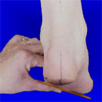
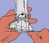
test that begins with the rearfoot in the position of subtalar congruity, with the midtarsal joint maximally pronated by applying an upward force equal to ground reactive force under the fifth metatarsal as Root described.
In my proposed Forefoot SERM-PERM Testing, the loaded plantar surface of the fifth metatarsal headserves as a new reference point to determine the position of the plantar surface of the first metatarsal head as it responds to testing.
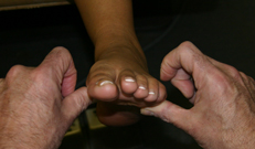
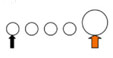
| If the first metatarsal head is below the fifth metatarsal after a test, it is recorded as plantarflexed. |
|
 |
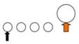
| If the first metatarsal head is on-line with the fifth metatarsal head after a test, it is recorded as on-line or parallel. |
|
 |
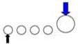
| If the first metatarsal head is above the fifth metatarsal after a test it is recorded as dorsiflexed. |
|
 |
| |
|
|
Forefoot SERM-PERM Testing
In Functional Foot Typing,4 Forefoot SERM-PERM Testing profiles every forefoot and classifies them into one of four (4) Forefoot Types. These are Rigid, Stable, Flexible and Flat. Each forefoot type has its own range of motion, closed chain pathology, predictable pain, overuse syndromes and pedal deformities, as well as treatment protocols that once diagnosed, enable skilled practitioners to apply focused kinetic and kinematic therapy that would be harmful to other foot types.
SERM Testing applies a downward force from above to the 1st metatarsal head and records the position as Plantarflexed or Dorsiflexed, PERM Testing applies an upward force from below the 1st metatarsal head and records the position as Plantarflexed, On-line or Dorsiflexed.
The Rigid Forefoot Type: In The Rigid Forefoot Type, the Forefoot SERM is Plantarflexed and the Forefoot PERM is Plantarflexed. |
|
|
The Stable Forefoot Type: In The Stable Forefoot Type, the Forefoot SERM is Plantarflexed and the Forefoot PERM is On-Line |
|
 |
The Flexible Forefoot Type: In the Flexible Forefoot Type, the Forefoot SERM is Plantarflexed and the Forefoot PERM is Dorsiflexed. |
|
|
The Flat Forefoot Type: In the Flat Forefoot Type, the Forefoot SERM is Dorsiflexed and the Forefoot PERM is Dorsiflexed. |
|
|
In summary, independent forefoot testing utilizing Forefoot SERM and PERM reduces the practitioners Rootian need to focus on the rearfoot when it comes to treatment. It draws attention to the forefoot as a source of pathology and a
location for treatment and rehabilitation and it is one of the keys to acculturating to Triplane Biomechanics. This quick, reproducible exam can be performed as part of The FFT Exam during the initial office visit in minutes and will revolutionize your practice.
References:
-
Harradine PD, Bevan LS: Gait dysfunction and podiatric therapy, British Journal of Podiatry, Feb 2003, 5-11
- Payne CB: The past, present, and future of podiatric biomechanics. JAPMA 88: 53, 1998, 77-81
- Kirby K: Are Root Biomechanics Dying? Podiatry Today, April 2009, Issue 4, 123-27
- Shavelson D: A closer look at neoteric biomechanics. Podiatry Today. Sept 2007;9, 234–241.
|
|