The Supine Biomechanical Evaluation
|
By Dennis Shavelson, DPM
Associate Editor |
Until the functional foot typing supine examination, biomechanical examination of the rearfoot and forefoot was performed with the patient in the prone position. The Subtalar Joint (STJ) was examined first and then the forefoot was "eyeballed" and measured by sighting beyond the rearfoot to the metatarsal head position. In order to upgrade the art, this author has turned the patient around and developed a biomechanical examination with the patient in the supine position and added a clearly defined forefoot examination opening up new doors in diagnosis, treatment, teaching and education of patients for podiatry and allied professions.
The Prone Biomechanical Evaluation
Root described the prone biomechanical examination in detail1 and it has since been interpreted by other examiners such as Palmer2. It attempts to determine Subtalar Neutral Position and Forefoot Position as an exact number of degrees claiming to be accurate and reproducible. In fact, the literature has shown prone examination to be inaccurate and not reproducible3,4. Furthermore, because the patient is facing away from the examiner and cannot visualize their feet, the prone bio eval is cold, impersonal and cannot be used for education. Even worse, since it is time consuming and needs an examination table that goes flat (180 degrees), its use on the initial examination, a critical time for introducing biomechanics to the patient is rare. More and more DPM’s fail to perform bio evals and a shoe clerk, a chiropractor or a podiatry assistant can find STJ Neutral position and using a box of foam or a scan generate custom orthotics as well as the average DPM. We have allowed biomechanics to become amateurish.
Clinicians deprive themselves of a valuable opportunity to professionally diagnose underlying biomechanical pathology and maybe more important, they are missing a critical moment to educate their patients about the importance of podiatry education and experience in diagnosis, casting, dispensing and monitoring custom foot orthotics during the initial exam.
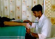
The Supine Biomechanical Evaluation
When the patient is turned around into supine position seated in a chair, the biomechanical evaluation becomes personal and allows the examiner to demonstrate to and educate the patient as they visualize the exam. It allows DPM’s renewed ability to separate from the less educated and skilled players dividing the biomechanical pie.
Separately profiling the rearfoot and forefoot utilizing supinatory and pronatory end range of motion (SERM and PERM) into one of four types, classify all feet into one of sixteen functional foot types serves as a neoteric foundation for podiatry to make a custom diagnosis and allow treatment of feet, the lower extremity and low back and many quality of life issues, professionally.5,6
Feet within each foot type share common characteristics that allow them to be batched in order to set up guidelines for care within that type. Since these characteristics are relative, depending on the education, the clinical experience and the skills of the examiner, patients receive different levels of care.
Podiatrists converted to supine biomechanical evaluation and foot typing once again provide more custom care, monitoring and professional assistance than others.
In summary, when compared to the prone bio eval, this neoteric supine examination is faster, more accurate, reproducible and requires more education and skill than its prone ancestor and because the patient can see their feet and the examiner when it is performed, the examination becomes educational and conducive to being performed during the initial office visit providing the opportunity for DPM’s to retake our place at the top of the biomechanical pyramid (see Table below).
Prone vs. Supine Bio Eval |
Prone Bio Eval
Needs a Flat
Examination Chair or Table |
Supine Bio Eval
Performed in
Any Chair or Table
|
Cold and Impersonal |
Patient Sees the Examiner and Their Feet
|
Tries to Take Exact Measurement |
Determines Rearfoot and
Forefoot Type Independently |
Not Educational |
Educational and Demonstrable |
Not Accurate or Reproducible |
Accurate and Reproducible |
20-30 Minutes |
4-6 Minutes |
Not Performed on the Initial Exam |
Easily Performed on the Initial Exam |
References:
-
Root ML, Orien WP, & Weed JH. Normal & abnormal function of the foot. (1977) Clinical Biomechanics Corporation, Los Angeles: Clinical Biomechanics
-
Palmer M, Epler M “Fundamentals of Musculoskelatal Assessment Techniques, 1998, Lippincott,
-
Chen Yan-xi, YU Guang-rong, Mei Jiong, Zhou Jia-qian. Assessment of subtalar joint neutral position: a cadaveric study. Chinese medical Journal, 2008, Vol.121 No. 8
-
Sobel E, Levitz, SJ. Reappraisal of the negative cast impression cast and subtalar neutral position. JAPMA:1997;87(1):30-34
-
Shavelson DE, A Closer Look at Neoteric Biomechanics; Podiatry Today: Volume 20; Sept 1, 2007; p.28-35
-
Shavelson, DE, Neoteric Biomechanics; Podiatry Management: September 2008; p. 122-126
|
|