| |
Robert Frykberg
DPM, MPH
PRESENT RI Editor
Diabetic Limb Salvage
|
Just when you think you’ve achieved success, the diabetic foot has a tendency to fool you. After many years of dealing with thousands of diabetic foot complications, I am always humbled by the challenges managing such disorders present to us. Even ostensibly straightforward problems can succumb to Murphy’s Law – if something can go wrong, it will, and often at the worst possible time! I think this really characterizes the diabetic foot and exemplifies why we must always be on our toes (forgive the pun) when managing patients with these difficult problems.
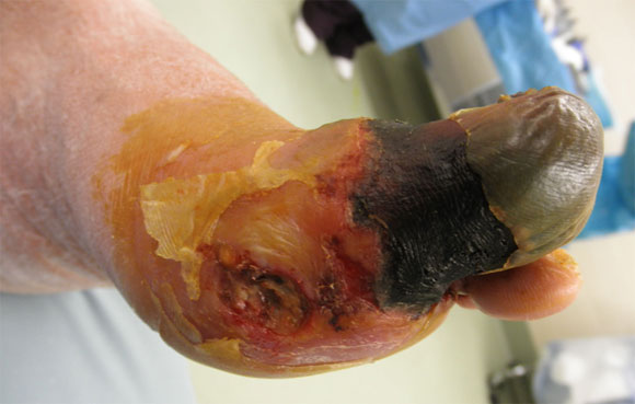
Going through my many cases, I found one that presented such a challenge, albeit not a horrible problem. Nonetheless, it seems to be a recurrent problem in our high risk patients. This patient, who we will refer to as Mr B., is a 58 year old type 2 diabetic man of 12 year’s duration. He presented to us upon referral from his Primary Care Physician with a gangrenous left great toe stemming from a shoe injury a month or so prior to presentation. He was clinically infected with cellulitis and a had a draining wound at the base of the toe. (Figure 1).
Figure 1
Gangrenous Left Hallux |
 |
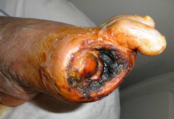
As is too often the case, his diabetes was not well controlled with a glycohemoglobin of 8.9%. White Blood Count (WBC) was at 10,000. Although neuropathic, he had non-palpable pedal pulses, monophasic Doppler signals, and an Ankle-Brachial Index (ABI) of 0.55, all confirming the presence of critical limb ischemia. He was promptly admitted and an open hallux amputation was performed to control the infection. As might be expected, this wound soon desiccated due to his ischemia. (Figure 2) Nonetheless, the infection was controlled (adjunctive antimicrobial therapy was also administered).
Figure 2
Post Hallux Amputation |
 |
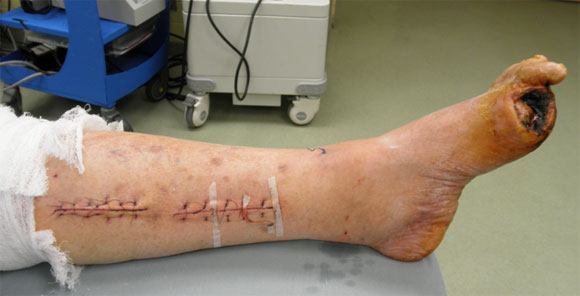
Based on angiography, a femoral-posterior tibial bypass was performed to improve perfusion as a limb salvage measure. (Figure 3)
Figure 3
Post Femoral-Posterior tibial bypass graft |
 |
This was a successful procedure as evidenced by the return of strong biphasic Doppler signals to the foot (although there were no granulations present at the prior hallux amputation site). Post revascularization transcutaneous oxygen measurement at the midfoot, however, revealed a pressure of only 35mmHg. Therefore, we performed a fairly classic transmetatarsal amputation (TMA), since we feared that any procedures distal to that level would be unsuccessful. (Figure 4)
Figure 4
Transmetatarsal amputation
| |